Regular Screening is Cervical Cancer Prevention

Screening is for healthy individuals with no symptoms. Cervical cancer screening helps to detect and treat cell changes at an early stage.

Regular testing can save your life and is the most effective method of preventing cervical cancer.

Screening

Cervical screening is not a test for cancer, it’s a test to help prevent cancer. The primary goal of screening is to identify precancerous lesions caused by HPV so they can be removed to prevent invasive cancers from developing.

A secondary goal is to find cervical cancers at an early stage when they can usually be treated successfully. Routine cervical screening has been shown to greatly reduce both the number of cervical cancer cases and deaths from the disease.

Prevention

Cervical cancer can often be prevented by having regular screenings with Pap tests and HPV tests to find any precancers and treat them. It can also be prevented by receiving the HPV vaccine. To help prevent cervical cancer it is recommended that girls receive HPV vaccination. HPV vaccination provides strong protection against new HPV infections.

HPV vaccination offers the most protection when given at ages 9-12. HPV vaccination is estimated to prevent up to 90% of HPV-related cancers.

Talk with your health care provider about the appropriate schedule for vaccination as it may vary based on many factors including age, gender and vaccine availability.

How is cervical cancer screening done?

Cervical cancer screening can be done in a medical office, a clinic, or a community health center. It is often done during a pelvic examination.

Research has shown that embarrassment, cultural barriers and fear are key underlying reasons to ignore cancer screening.

The earlier HPV‑induced‑ or p16/Ki‑67‑positive cervical lesions are detected and treated, the better. It could save your life!

Typical Procedure

While a woman lies on an exam table, a health care professional inserts an instrument called a speculum into her vagina to widen it so that the upper portion of the vagina and the cervix can be seen.

This procedure also allows the health care professional to take a sample of cervical cells. The cells are taken with a wooden or plastic scraper and/or a cervical brush and placed in a vial of liquid preservative.

The home swab offered in some countries is a simple way for women to do the test themselves. These home kits give thousands of women another option to keep up to date with their screening.

Laboratory Testing

The sample is sent to a laboratory where the cells are tested for the presence of high-risk types of HPV and / or examined under a microscope with an automated liquid-based Pap cytology test. When both tests are done using the same sample, this is referred to as “co-testing”.

With the introduction of the liquid-based Papanicolaou test, high-risk HPV testing, and computer-assisted screening, the cytology laboratory is at the forefront of efforts to improve screening for the provision of better patient care.

The Bethesda System

The Bethesda System considers abnormalities of squamous cells and glandular cells separately. These two cell types meet at a place called the transformation zone. The exact location of the transformation zone changes as you get older and if you give birth.

Squamous cell abnormalities are divided into the following categories, ranging from the mildest to the most severe:

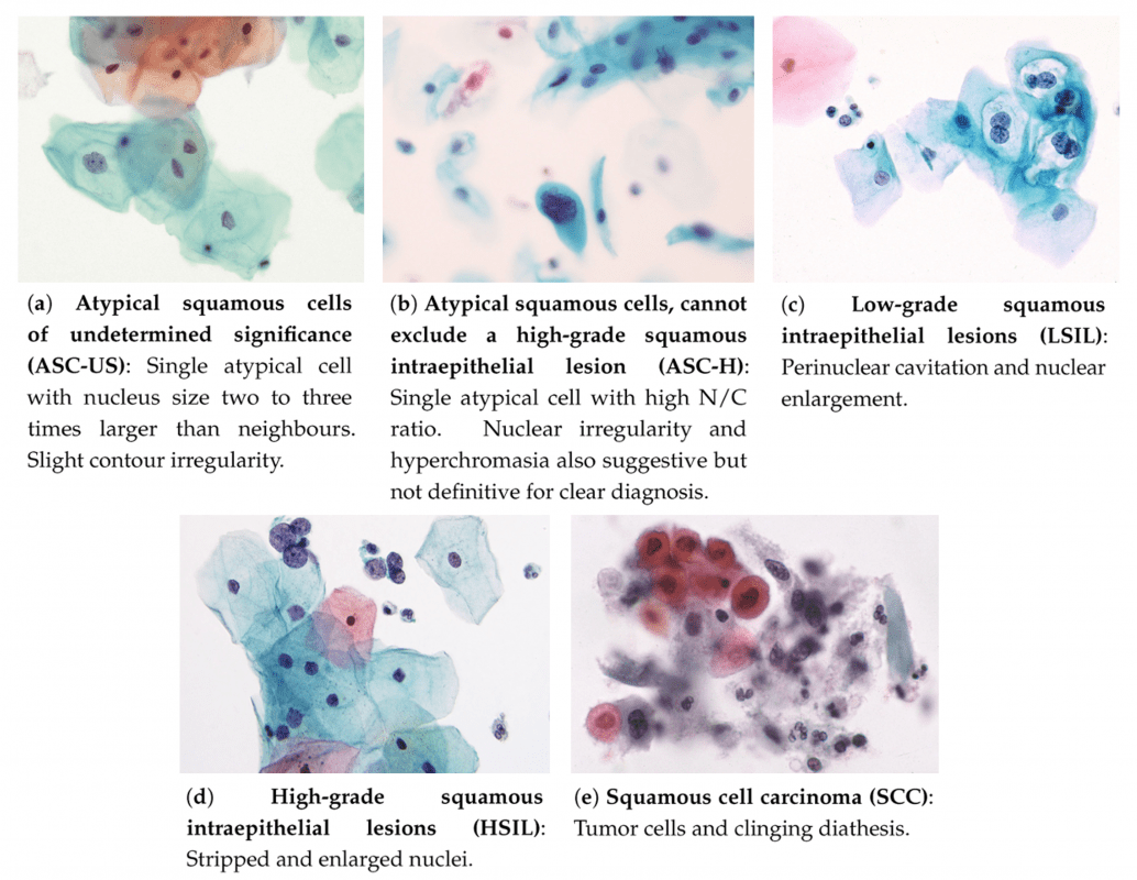
Examples of atypical squamous cells on liquid-based cytology (LBC). From: Nayar, R.; Wilbur, D. The Bethesda System for Reporting Cervical Cytology: Definitions, Criteria, and Explanatory Notes, 3rd ed.; Springer International Publishing, 2015 a.

Squamous cell abnormalities

  • Atypical squamous cells (ASC) are the most common abnormal finding in Pap tests. The Bethesda System divides this category into two groups, ASC‑US and ASC‑H:
    • ASC-US: atypical squamous cells of undetermined significance. The cells do not appear completely normal, but the cause is unclear. The changes may be related to an HPV infection, but they can also be caused by other factors.
    • ASC-H: atypical squamous cells, cannot exclude a high-grade squamous intraepithelial lesion. ASC-H lesions may be at higher risk of being precancerous than ASC-US lesions.
  • Low-grade squamous intraepithelial lesions (LSILs) are considered mild abnormalities caused by HPV infection. LSILs often return to normal as the immune system controls the infection, especially in younger women.
  • High-grade squamous intraepithelial lesions (HSILs) are more severe abnormalities that have a higher likelihood of progressing to cancer if left untreated.
  • Carcinoma in situ (CIS) refers to severely abnormal cells that resemble cancer cells but remain on the surface of the cervix and have not invaded more deeply or spread beyond the cervix.
  • Squamous cell carcinoma is cervical cancer. The abnormal squamous cells have invaded more deeply into the cervix or into other tissues or organs. In a well-screened population, such as that in the United States, a finding of cancer during cervical screening is extremely rare.

Glandular cell abnormalities

Atypical glandular cells (AGC) on cervical cytology usually originate from the glandular epithelium of the endocervix or endometrium. They are a less common finding than abnormal squamous cells.

Glandular cell abnormalities describe abnormal changes that occur in the glandular tissues of the cervix. The Bethesda system divides these abnormalities into the following categories:

  • Atypical glandular cells (AGC), meaning the glandular cells do not appear normal, but doctors are uncertain about what the cell changes mean.
  • Endocervical adenocarcinoma in situ (AIS), meaning that severely abnormal cells are found but have not spread beyond the glandular tissue of the cervix.
  • Adenocarcinoma includes not only cancer of the endocervical canal itself but also, in some cases, endometrial, extrauterine, and other cancers.

Munich Nomenclature

Different classification systems for Pap smear samples have been internationally used. Germany applies the Munich nomenclature for Pap smear evaluation. The Munich nomenclature was established in 1975 as a modification of Papanicolaou’s classification.

This modification was necessary to meet the international requirements of a descriptive classification. In 2015 the Munich III nomenclature has been established as the only system officially used in Germany.

With Munich III, new subgroups were created to categorize different grads of dysplasia. Unclear findings that are neither clearly reactive nor meet certain criteria of dysplasia are now marked. Munich III further differentiates between squamous, epithelial, and glandular cells.

Furthermore, the Munich III system is attempting to make cytological findings transferrable to the internationally more commonly used The Bethesda System. This offers the opportunity to compare them with international studies.

Screening Methods

For many years, cytology-based screening, known as the Pap test or Pap smear, was the only method of screening. Its use reduced cervical cancer incidence and deaths in countries where screening is common. However, with the advent of the ability to test for HPV, cervical cancer screening now includes three approaches:

HPV testing, which looks for the presence of high-risk HPV types in cervical cells, Pap testing and HPV/Pap co-testing, which checks the same cell sample for both high-risk HPV types and cervical cell changes.

HPV Test and Pap Test

The HPV test and the Pap test are two different cervical cancer screening tests. Screening means checking for a disease or for changes that may develop into disease before there are symptoms.

Women need routine cervical cancer screenings even if they feel fine. Screening can help find changes in cervical cells so you can receive the proper follow-up care and treatment you need to stay healthy.

  • The HPV test checks cells for infection with high-risk HPV types.
  • The Pap test (also called a Pap smear or cervical cytology) collects cervical cells and looks at them for changes caused by HPV that may—if left untreated—turn into cervical cancer. It can also detect cervical cancer cells. A Pap test also sometimes finds conditions that are not cancer, such as infection or inflammation.
  • The HPV/Pap co-test uses a Pap test and HPV test together to check for both high-risk HPV and cervical cell changes.

CINtec® PLUS

CINtec® PLUS Cytology is a triage test for cervical cancer screening, and the only test that uses dual-biomarker technology to simultaneously detect p16 and Ki-67 to provide a strong indicator of the presence of transforming HPV infections.

CINtec® PLUS Cytology objectively identifies transforming HPV infections in women with abnormal Pap cytology results and HPV-positive cervical cancer screening results, allowing laboratory professionals to help physicians be certain they can recommend follow-up for women who will benefit most from colposcopy.

  • The test indicates oncogenically transforming HPV infections, and thus eliminates equivocation over morphologic abnormalities that are not pre-cancerous. Very good overall clinical accuracy has been demonstrated in key cervical cancer screening scenarios.
  • Overall, p16/Ki‑67 dual staining has the potential to significantly reduce the number of women referred for confirmatory colposcopy.

Cervical Erosions

Cervical erosion is sometimes called cervical eversion, cervical ectropy, or cervical ectropion. It is a common condition in women who are in their childbearing years and usually nothing to worry about. It is not a sign of another health problem, like cervical cancer. But if you have symptoms that bother you, there are treatments your doctor can try.

Cervical ectropion happens when cells that line the inside of your cervix grow on the outside. These cells are redder and are more sensitive than the cells typically on the outside, which is why they may cause symptoms, like bleeding and discharge, for some women.

GET ACTIVE